


Mark Zuckerberg says Meta wearables that read brain signals are coming soon
SOURCE: COINTELEGRAPH.COM
APR 19, 2024
This new wearable can image a bodily organ as well as an ultrasound
SOURCE: HTTPS://INTERESTINGENGINEERING.COM/
NOV 16, 2023
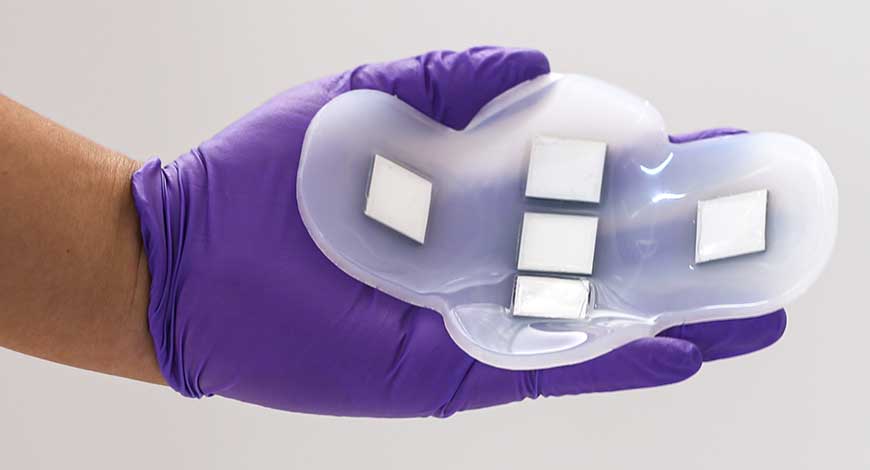
Researchers at MIT have created a wearable ultrasound monitor that resembles a patch and can image internal organs without the use of medical tools.
“This technology is versatile and can be used not only on the bladder but any deep tissue of the body. It’s a novel platform that can do identification and characterization of many of the diseases that we carry in our body,” said Canan Dagdeviren, an associate professor in MIT’s Media Lab and the senior author of the study.
The scientists decided to begin by testing the patch on the bladder. The decision to concentrate on this body organ for the first demonstration was made, in part, because Dagdeviren's younger brother was recently diagnosed with kidney cancer.
The surgical removal of one of his kidneys left him unable to completely empty his bladder. Dagdeviren questioned whether individuals with kidney or bladder issues may benefit from an ultrasound monitor that would indicate the amount of fluid in the bladder.
Additionally, the researchers wanted to create a wearable substitute that patients may use in their homes, reducing visits to health care facilities.
“Millions of people are suffering from bladder dysfunction and related diseases, and not surprisingly, bladder volume monitoring is an effective way to assess your kidney health and wellness,” she said
The novel wearable is comprised of a silicone rubber patch that is flexible and soft to the touch. It is embedded with five ultrasonic arrays arranged in the shape of a cross and created by the researchers using a novel piezoelectric material. The polymer that comprises the patch is naturally adhesive and sticks to the skin softly: pants or leggings can easily hold it in place once it's applied to the skin and it can just as easily be removed.
Further tests conducted by the researchers demonstrated that the novel patch could record images that were on par with those obtained using conventional ultrasonic probe.
Twenty participants with a range of body mass indices were enrolled in the trials of the patch. First, the bladders of the subjects were photographed full, then partially empty, and finally entirely empty. The novel patch was demonstrated to produce high-quality images comparable to those acquired using conventional ultrasound, and the ultrasonic arrays were effective on all participants, irrespective of their body mass index.
The same type of ultrasound machine used in medical imaging centers was connected to the researchers' ultrasonic arrays so they could view the images. To examine the photos, though, the MIT team is currently developing a small portable gadget that resembles a smartphone.
“For whatever organ that we need to visualize, we go back to the first step, select the right materials, come up with the right device design and then fabricate everything accordingly,” said Dagdeviren.
The MIT group now also plans to create ultrasonic imaging tools that might be used to explore other human organs including the liver, pancreas, and ovaries.
The study is published in the journal Nature Electronics.
LATEST NEWS
WHAT'S TRENDING


Data Science
5 Imaginative Data Science Projects That Can Make Your Portfolio Stand Out
OCT 05, 2022

SOURCE: COINTELEGRAPH.COM
APR 19, 2024
SOURCE: HTTPS://WWW.AZOSENSORS.COM/NEWS
AUG 28, 2023
SOURCE: WWW.WHATGADGET.NET
JUL 11, 2023
SOURCE: HTTPS://WWW.THEHINDU.COM/SCI-TECH/TECHNOLOGY/INDIAS-WEARABLE-MARKET-GREW-47-IN-2022-BOAT-MAINTAINS-LEAD-REPORT/ARTICLE66493121.ECE
JUN 30, 2023


SOURCE: THECONVERSATION.COM
OCT 16, 2022